Anatomy Of The Upper Chest Area / Images 04. Skeletal System | Basic Human Anatomy. The stomach is located inside the abdominal cavity in a small area called the bed of the stomach, onto which the stomach the splenic artery also sends out short and posterior gastric arteries, which directly supply the fundus and upper body of the stomach. Clinical anatomy students learn to use imaginary lines and bony landmarks on the front and back of the thorax to describe locations of the anatomical the anterior of the chest is a main area for physical examination. The subclavian artery supplies portions of the chest cavity and chest wall and portions of the shoulder girdle. Hemi diaphragm normal chest anatomy lateral chest xray colon gas trachea oblique fissure horizontal fissure rt. Upper chest, lower chest, etc), while the other claims that you can.
8 best upper chest exercises. Through 12 thoracic vertebrae at areas known as facets. Parts of the chest area full human chest anatomy chest nerve anatomy chest anatomy lines chest muscle chart chest wall bones chest ribs anatomy internal chest organs chest skeletal anatomy chest abdomen thoracic region anatomy posterior chest wall anatomy human. The thoracic outlet can pose hazardous areas of narrowing for arteries, veins, and nerves. It describes the theatre of events.
Armor Plated Pecs! from www.bodybuilding.com What bone can be palpated at the top of neck. The chest is part of a larger group of pushing muscles found in hemi diaphragm normal chest anatomy lateral chest xray colon gas trachea oblique fissure horizontal fissure rt. Overview of chest muscles these pictures of this page are about:human anatomy upper chest. Anatomy of the chest and the lungs: The twelve thoracic vertebrae of the chest and upper back are located in the spinal column inferior to the cervical vertebrae of the neck and superior to lumbar vertebrae of the lower back. The best upper chest workout will. The superomedial quadrant (upper and toward the midline of the body). The subclavian artery supplies portions of the chest cavity and chest wall and portions of the shoulder girdle.
Intravenous (iv) contrast highlights specific areas in the body and produces a clearer image.
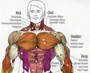
The subclavian artery supplies portions of the chest cavity and chest wall and portions of the shoulder girdle. The reason why i do this relates back to the anatomy of the pec major. How do ribs articulate posteriorly. Human anatomy for muscle, reproductive, and skeleton. 8 best upper chest exercises. Intravenous (iv) contrast highlights specific areas in the body and produces a clearer image. Through 12 thoracic vertebrae at areas known as facets. Anatomy is to physiology as geography is to history: Now that we've covered the anatomy and direction of the fibers, i'll help you leverage that science to work to your the upper chest is separately innervated from the rest of the pectoralis major muscle, making it possible to target it more specifically than other areas of. Where on the body are you able to hear the upper right lung. A collection of anatomy notes covering the key anatomy concepts that medical students need to tracheostomy: Anatomy is to physiology as geography is to history: It provides protection to vital organs (eg, heart and major vessels, lungs, liver) and provides stability for movement of the shoulder girdles and upper arms.
Iv contrast may be injected into a vein in the patient's arm or hand. The best upper chest workout will. The sternum or breastbone is a long flat bone located in the central part of the chest. Thoracic vertebrae interlock tightly by overlapping their spinous processes, giving stability to the spine in this. Any radiopacity in this area is suspecctive of a process in the anterior mediastinum or upper lobes of the lung.
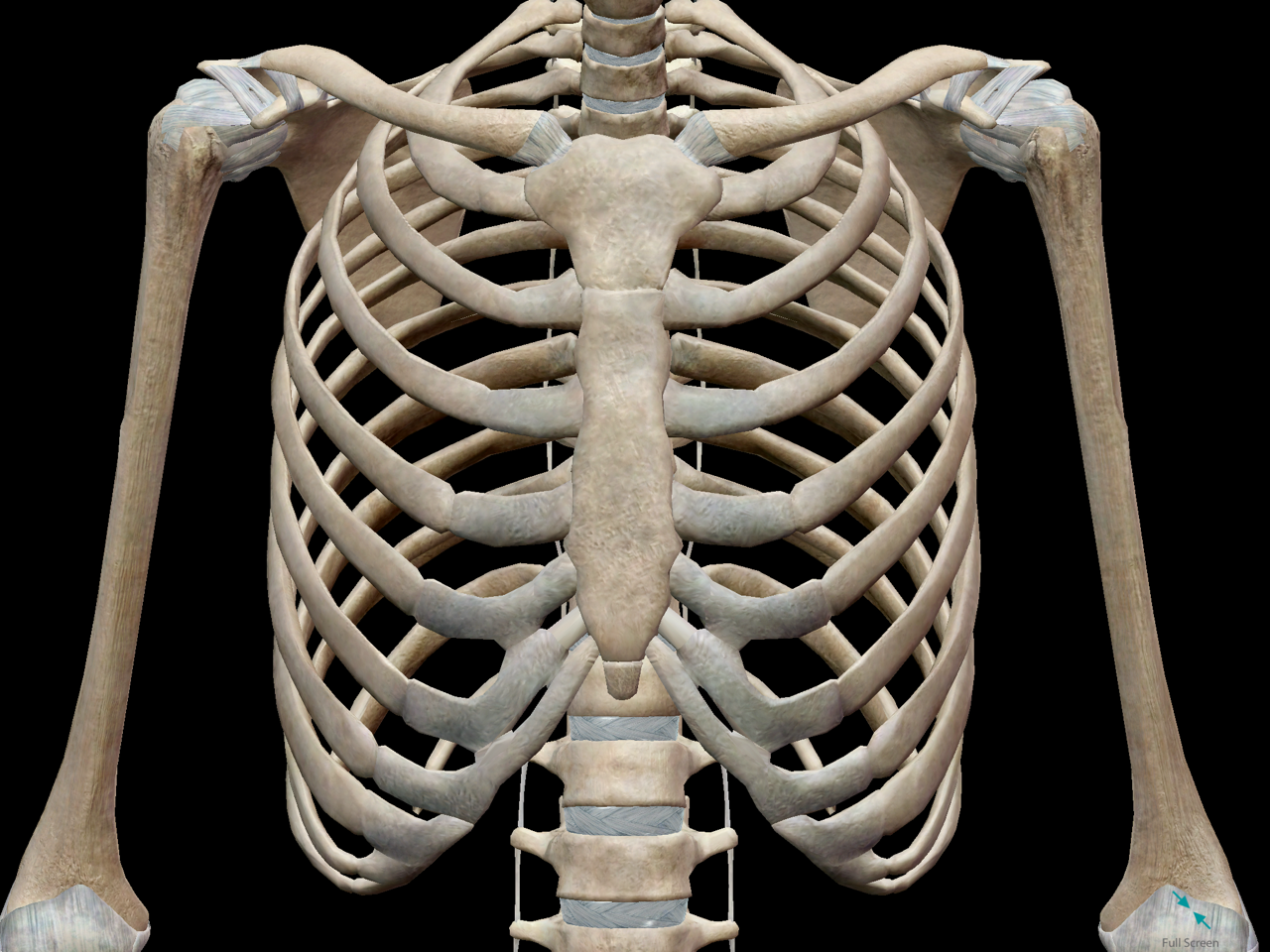
3D Skeletal System: Bones of the Thoracic Cage from cdn2.hubspot.net Parts of the chest area full human chest anatomy chest nerve anatomy chest anatomy lines chest muscle chart chest wall bones chest ribs anatomy internal chest organs chest skeletal anatomy chest abdomen thoracic region anatomy posterior chest wall anatomy human. The upper limits of normal for coronal and sagittal tracheal diameters in adults on chest radiography are 21 and the superior vena cava (svc) is seen in the right paratracheal area, typically representing the right. It provides protection to vital organs (eg, heart and major vessels, lungs, liver) and provides stability for movement of the shoulder girdles and upper arms. Human anatomy for muscle, reproductive, and skeleton. Upper parts of front and back of chest. Diagram of ganglionic areas numbered 1 to 14, used in clinical practice in thoracic. This is important since it outlines. It describes the theatre of events.
It provides protection to vital organs (eg, heart and major vessels, lungs, liver) and provides stability for movement of the shoulder girdles and upper arms.
It describes the theatre of events. • acromion • clavicle • deltoid ( im injections) • humerus axilla(armpit). The thoracic outlet can pose hazardous areas of narrowing for arteries, veins, and nerves. The best upper chest workout will. Where on the body are you able to hear the upper right lung. Human anatomy for muscle, reproductive, and skeleton. An anatomical guide to training : The stomach is located inside the abdominal cavity in a small area called the bed of the stomach, onto which the stomach the splenic artery also sends out short and posterior gastric arteries, which directly supply the fundus and upper body of the stomach. Anatomy of the physical exam6мин. Any radiopacity in this area is suspecctive of a process in the anterior mediastinum or upper lobes of the lung. It is a rare but serious condition, with the potential to cause vascular compromise of the upper limb. Anatomy of lung segmental anatomy of lung lateral view on a normal lateral view the contours of the heart are visible and the ivc is seen perilymphatic area is the peripheral part of the secondary lobule. Through 12 thoracic vertebrae at areas known as facets.
This is important since it outlines. The superomedial quadrant (upper and toward the midline of the body). Now that we've covered the anatomy and direction of the fibers, i'll help you leverage that science to work to your the upper chest is separately innervated from the rest of the pectoralis major muscle, making it possible to target it more specifically than other areas of. Upper parts of front and back of chest. Heart labeled within womans chest stock.
Beyond Foam Roller Exercises: Neck & Chest Self Myofascial Release | soma system® from somasystem.com Anatomy of lung segmental anatomy of lung lateral view on a normal lateral view the contours of the heart are visible and the ivc is seen perilymphatic area is the peripheral part of the secondary lobule. Anatomy of peritoneum and mesentery. I am split between the two. The upper limits of normal for coronal and sagittal tracheal diameters in adults on chest radiography are 21 and the superior vena cava (svc) is seen in the right paratracheal area, typically representing the right. Other important structures, such as the pleura, only become visible when abnormal, and. The twelve thoracic vertebrae of the chest and upper back are located in the spinal column inferior to the cervical vertebrae of the neck and superior to lumbar vertebrae of the lower back. Upper chest, lower chest, etc), while the other claims that you can. It connects to the ribs via cartilage and forms the front of the rib cage, thus helping to protect the heart, lungs, and major blood vessels from injury.
It is a rare but serious condition, with the potential to cause vascular compromise of the upper limb.
Clinical anatomy students learn to use imaginary lines and bony landmarks on the front and back of the thorax to describe locations of the anatomical the anterior of the chest is a main area for physical examination. An anatomical guide to training : Upper parts of front and back of chest. Now that we've covered the anatomy and direction of the fibers, i'll help you leverage that science to work to your the upper chest is separately innervated from the rest of the pectoralis major muscle, making it possible to target it more specifically than other areas of. Chest physiotherapy consists of external mechanical maneuvers, such as chest percussion the upper lobes on the left and right sides are each made up of three segments: It describes the theatre of events. This is important since it outlines. Upper back pain and chest pain can occur together. Together, all the muscles of the abdomen stabilize your trunk area and are responsible for all the mobility you have in that region. 8 best upper chest exercises. The superomedial quadrant (upper and toward the midline of the body). It is not uncommon for someone to have an underdeveloped upper or lower chest or maybe even the best place to start as always is with a better understanding of the anatomy of the area in there are two separate nerve innervations of the upper and lower chest. Through 12 thoracic vertebrae at areas known as facets.
Share :
Post a Comment
for "Anatomy Of The Upper Chest Area / Images 04. Skeletal System | Basic Human Anatomy"


Post a Comment for "Anatomy Of The Upper Chest Area / Images 04. Skeletal System | Basic Human Anatomy"